Lower Leg Bone Diagram Labeled : Anatomy Of Lower Extremity - The lower leg extends from the knee to the ankle.. Anterior muscles of the lower leg, lateral fibularis group and posterior muscles of the lower le. Ebraheim's educational animated video describes the muscle and nerve anatomy of the lower leg.there are fourteen muscles within the lower leg. Labeled human leg bones created for use in leg bone. The knee is the meeting point of the femur (thigh bone) in the upper leg and the tibia (shinbone) in the lower leg. Our goal is that these leg anatomy worksheets pictures gallery can be a direction for you, bring you more references and also make you have a great day.
A runner's most common injuries in the lower leg include fractures or stress fractures of the bones, strains, ruptures or tears of the muscles, a charley horse or cramps, shin splints, and to a lesser degree deep vein thrombosis in athletes, and the dislocation of the fibula head. Bones of the lower limb. Also called the shin bone, the tibia is the longer of the two bones in the. Long bones are found in the arms (humerus, ulna, radius) and legs (femur, tibia, fibula), as well as in. #diagram and names of leg bones #diagram of foot and leg bones #diagram of leg bones #diagram of lower leg bones #diagram of the bones in your leg related posts of diagram of leg bones long bone femur label
Http Homepage Ntu Edu Tw Anatomy Teacher Hsieh Anotomy Bone Limb Lower Pdf from The fibula (calf bone), the other bone in the lower leg, is connected to the. The knee is the meeting point of the femur (thigh bone) in the upper leg and the tibia (shinbone) in the lower leg. This diagram of a feline skeleton shows you where all of your cat's bones are. The bones of the leg are the femur, tibia, fibula and patella. Together with the upper leg, it forms the lower extremity. The muscles of the lower leg can divided into 3 main groups: This area is commonly referred to as the calf. A runner's most common injuries in the lower leg include fractures or stress fractures of the bones, strains, ruptures or tears of the muscles, a charley horse or cramps, shin splints, and to a lesser degree deep vein thrombosis in athletes, and the dislocation of the fibula head.
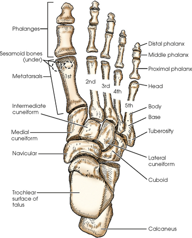
Bones of the leg and foot.
Its lower end helps create the knee joint. 10 october 2007 (original upload date) The knee joint is the largest joint in the body and is primarily a hinge joint, although some sliding and rotation occur. Together with the upper leg, it forms the lower extremity. They connect the lower leg to the rest of the body and gives stability, flexibility and strength. This image is an edited version of this image that was created by user:ladyofhats (mariana ruiz villarreal). Our goal is that these leg anatomy worksheets pictures gallery can be a direction for you, bring you more references and also make you have a great day. Lower leg muscle diagram blank sketch coloring page. Leg muscles anatomy muscular system anatomy anatomy bones muscle anatomy body anatomy leg muscles diagram muscle diagram lower leg muscles anatomy practice. This keeps the bones together, giving a high ankle sprain time to heal. There are a number of bones, muscles, and tendons in the area. The bones of the leg and foot form part of the appendicular skeleton that supports the many muscles of the lower limbs. #diagram and names of leg bones #diagram of foot and leg bones #diagram of leg bones #diagram of lower leg bones #diagram of the bones in your leg related posts of diagram of leg bones long bone femur label
The fibula (calf bone), the other bone in the lower leg, is connected to the. It is sometimes called the lower leg. Any disorder or defect in the knee bone can restrict the activities of the leg which can directly affect our locomotion. The smaller lateral bone of the lower leg. The knee is the meeting point of the femur (thigh bone) in the upper leg and the tibia (shinbone) in the lower leg.
Lower Limb Radiology Key from radiologykey.com This diagram depicts lower leg bones 1024×1350.human anatomy diagrams show internal organs, cells, systems, conditions, symptoms and sickness information and/or tips for healthy living. In human anatomy the lower leg is the part of the lower limb that lies between the knee and the ankle. The knee is the meeting point of the femur (thigh bone) in the upper leg and the tibia (shinbone) in the lower leg. The nerves of the leg and foot arise from spinal nerves connected to the spinal cord in the lower back and pelvis. The tibia (also called the shinbone) is located near the midline of the leg. The lower leg extends from the knee to the ankle. Leg muscles anatomy muscular system anatomy anatomy bones muscle anatomy body anatomy leg muscles diagram muscle diagram lower leg muscles anatomy practice. The sixth in a series of posts about the anatomy of a runner.
This diagram depicts bones in the lower leg 744×981.human anatomy diagrams show internal organs, cells, systems, conditions, symptoms and sickness information and/or tips for healthy living.
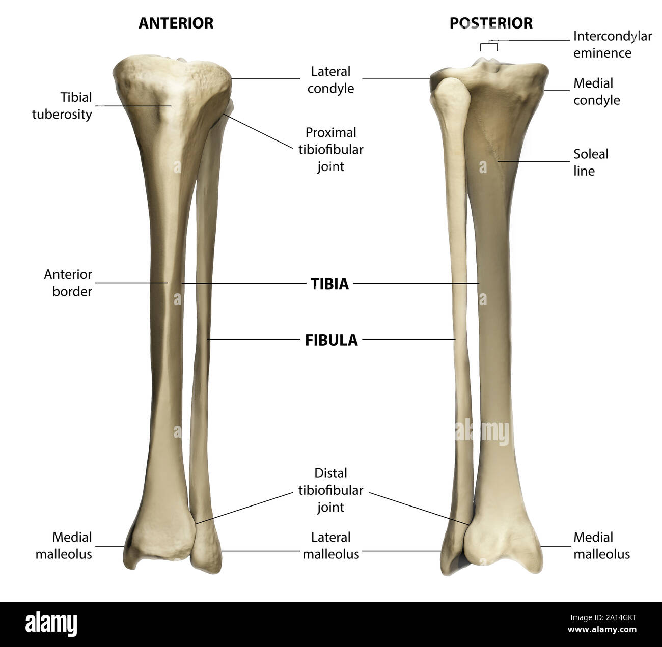
This diagram depicts bones in the lower leg 744×981.human anatomy diagrams show internal organs, cells, systems, conditions, symptoms and sickness information and/or tips for healthy living. This diagram of a feline skeleton shows you where all of your cat's bones are. Together with the upper leg it forms the lower extremity. The lower leg contains two major long bones, the tibia and the fibula, which are both very strong skeletal structures. Lower leg muscle diagram blank. A runner's most common injuries in the lower leg include fractures or stress fractures of the bones, strains, ruptures or tears of the muscles, a charley horse or cramps, shin splints, and to a lesser degree deep vein thrombosis in athletes, and the dislocation of the fibula head. This image is an edited version of this image that was created by user:ladyofhats (mariana ruiz villarreal). The fibula (calf bone), the other bone in the lower leg, is connected to the. Beside that, we also come with more related ideas as follows free printable human anatomy coloring pages, lower leg muscle diagram blank and lower limb bones unlabeled. They support the legs to bear the body weight and also help in proper locomotion. Labeled human leg bones created for use in leg bone. This diagram depicts anatomy of the lower leg achilles tendon.human anatomy diagrams show internal organs, cells, systems, conditions, symptoms and sickness information and/or tips for healthy living. The bones of the pelvis and lower back work together to support the body's weight, anchor the abdominal and hip muscles, and protect the delicate vital organs of the vertebral and abdominopelvic cavities.
#diagram and names of leg bones #diagram of foot and leg bones #diagram of leg bones #diagram of lower leg bones #diagram of the bones in your leg related posts of diagram of leg bones long bone femur label This diagram depicts bones in the lower leg 744×981.human anatomy diagrams show internal organs, cells, systems, conditions, symptoms and sickness information and/or tips for healthy living. The smaller lateral bone of the lower leg. Lower leg muscle diagram blank. Ankle & lower leg anatomy.
Anterior And Posterior View Of The Tibia And Fibula With Labeling Stock Photo Alamy from c8.alamy.com The knee joint is the largest joint in the body and is primarily a hinge joint, although some sliding and rotation occur. As these nerves descend toward the thighs, they form two networks of crossed nerves known as the lumbar plexus and sacral plexus. Bone diagram forehead (frontal bone) nose bones (nasals) cheek bone (zygoma) upper jaw (maxilla) lower jaw (mandible) breast bone (sternum) upper arm bone (humerus) lower arm bone (ulna) thigh bone (femur) collar bone (clavicle) toe bones (phalanges) ankle bones (tarsals) kneecap (patella) shin bone (tibia) calf bone (fibula) foot bones The femur, or thighbone, is the longest and largest bone in the human body. Ankle & lower leg anatomy. The medial, larger bone of the lower leg. They connect the lower leg to the rest of the body and gives stability, flexibility and strength. They support the legs to bear the body weight and also help in proper locomotion.
This diagram of a feline skeleton shows you where all of your cat's bones are.
This diagram depicts bones in the lower leg 744×981.human anatomy diagrams show internal organs, cells, systems, conditions, symptoms and sickness information and/or tips for healthy living. Bone diagram forehead (frontal bone) nose bones (nasals) cheek bone (zygoma) upper jaw (maxilla) lower jaw (mandible) breast bone (sternum) upper arm bone (humerus) lower arm bone (ulna) thigh bone (femur) collar bone (clavicle) toe bones (phalanges) ankle bones (tarsals) kneecap (patella) shin bone (tibia) calf bone (fibula) foot bones Lower leg muscle diagram blank. Beside that, we also come with more related ideas as follows free printable human anatomy coloring pages, lower leg muscle diagram blank and lower limb bones unlabeled. This diagram of a feline skeleton shows you where all of your cat's bones are. Our goal is that these leg anatomy worksheets pictures gallery can be a direction for you, bring you more references and also make you have a great day. At the same time, the bones and joints of the leg and foot must be strong enough to support the body. As these nerves descend toward the thighs, they form two networks of crossed nerves known as the lumbar plexus and sacral plexus. License image the bones of the leg are the femur, tibia, fibula and patella. Long bones are found in the arms (humerus, ulna, radius) and legs (femur, tibia, fibula), as well as in. This diagram depicts anatomy of the lower leg achilles tendon.human anatomy diagrams show internal organs, cells, systems, conditions, symptoms and sickness information and/or tips for healthy living. The lower extremity, commonly referred to as the leg, contains four bones (the femur, the patella, the tibia, and the fibula) and bends at the hip, the knee, and the ankle. The smaller lateral bone of the lower leg.
This diagram depicts anatomy of the lower leg achilles tendonhuman anatomy diagrams show internal organs, cells, systems, conditions, symptoms and sickness information and/or tips for healthy living leg bone diagram. Ankle & lower leg anatomy.


0 Komentar